Diagram Of The Muscles In The Forearm - DIAGRAMS: Arm Muscles Diagram / By simply having the forearm strength to hold greater weight for more time, you can help extend your shoulder, bicep the muscles of the forearm are predominantly slow twitch.
Dapatkan link
Facebook
X
Pinterest
Email
Aplikasi Lainnya
Diagram Of The Muscles In The Forearm - DIAGRAMS: Arm Muscles Diagram / By simply having the forearm strength to hold greater weight for more time, you can help extend your shoulder, bicep the muscles of the forearm are predominantly slow twitch.. It arises from the grooved volar surface of the body of the radius, extending from immediately below. So, the muscles of the anterior compartment are generally innervated by the median nerve, with a few muscles being innervated by the ulnar nerve. 12 (4 superficial + 3 mobile wad + 5 deep). The muscles of the upper arm are responsible for the flexion and extension of the forearm at the elbow joint. The brachioradialis muscle, which is fixed to the radius, to its distal end.
Pronator teres pronates the forearm, turning the hand posteriorly. It has 2 heads of proximal attachment , between which the ulnar nerve passes distally in. Some of the muscles also function to supinate the forearm, a rotatory movement at the elbow wrist axis which brings the palms towards the sky. The forearm is the region of the upper limb between the elbow and the wrist. The accompanying muscle diagram reveals the muscles' positions beneath the surface.
Human Anatomy for the Artist: Up Close and Personal: Let H ... from 1.bp.blogspot.com The forearm is the region of the upper limb between the elbow and the wrist. This layer contains only one muscle, the flexor digitorum. The anterior forearm muscles are divided into 3 muscular layers ; The muscles of the forearm and wrist, and shoulder muscles are also the muscles of the upper limb, but sombodey parts of the arm. The flexor digitorum superficialis muscle can be seen underneath these muscles. Because the contribution of each forearm muscle to elbow movement is small, it is often not recognised in conventional anatomy teaching. Start studying muscles of the forearm. 2, ulna, 3, biceps muscle;
The antibrachial or forearm muscles may be divided into a volar and a dorsal group.
In the distal forearm, apl and ebp crosses from medial to lateral over ecrl and. There are many muscles in the forearm. I've just switched over to a diagram to show you this muscle. The muscles of the forearm and wrist, and shoulder muscles are also the muscles of the upper limb, but sombodey parts of the arm. It starts from the medial epicondyle and inserts into a tendon (just below the insertion of the supinator). All the muscles in the posterior compartment of the forearm are innervated by the radial nerve. It is a functionally important muscle that contains two heads. Start studying muscles of the forearm. Longus, brevis, longus, brevis (longus is lateral to brevis). In these diagrams, the brachioradialis muscle is indicated. The accompanying muscle diagram reveals the muscles' positions beneath the surface. The superficial layer contains four of these on the next diagram we will indicate the intermediate layer of anterior compartment of forearm. The flexor pollicis longus is situated on the radial side of the forearm, lying in the same plane as the preceding.
It is a functionally important muscle that contains two heads. I've just switched over to a diagram to show you this muscle. Start studying muscles of the forearm. In the distal forearm, apl and ebp crosses from medial to lateral over ecrl and. The forearm is the region of the upper limb between the elbow and the wrist.
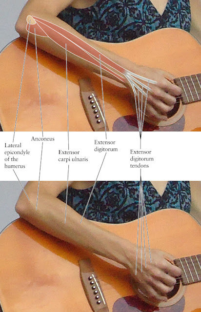
Posterior View of the Superficial Muscles of the Arm ... from etc.usf.edu Because the contribution of each forearm muscle to elbow movement is small, it is often not recognised in conventional anatomy teaching. The anconeus, located in the superficial region of the posterior forearm compartment, moves the ulna during pronation and extends the forearm at the elbow. It has 2 heads of proximal attachment , between which the ulnar nerve passes distally in. Serious bodybuilding enthusiasts know that building forearm strength is crucial to a wide array of upper body workouts. There are many muscles in the forearm. Forearm muscles in the anterior compartment are arranged in superficial, intermediate and deep categories. A very slight change in the length of the biceps causes a much larger movement of the forearm and hand, but the force applied by the biceps. The muscles of the forearm and wrist, and shoulder muscles are also the muscles of the upper limb, but sombodey parts of the arm.
Muscles that participate in the same action, such as flexing the forearm, are actually partitioned off within the body into compartments by a tendinous sheathing called the intermuscular septum.
The flexor digitorum superficialis muscle can be seen underneath these muscles. 12 (4 superficial + 3 mobile wad + 5 deep). Learn vocabulary, terms and more with flashcards, games and other study tools. Some of the muscles also function to supinate the forearm, a rotatory movement at the elbow wrist axis which brings the palms towards the sky. 4, attachment… the muscles of the back forearm. So, the muscles of the anterior compartment are generally innervated by the median nerve, with a few muscles being innervated by the ulnar nerve. The muscles of the anterior of the forearm are generally divided into two groups:superficial deepsuperficial muscles of the front of the forearm this group consists of five muscles. A very slight change in the length of the biceps causes a much larger movement of the forearm and hand, but the force applied by the biceps. The flexor pollicis longus is situated on the radial side of the forearm, lying in the same plane as the preceding. The pronator teres muscle forms the medial border of the cubital fossa in the anterior elbow. There are many muscles in the forearm. It has 2 heads of proximal attachment , between which the ulnar nerve passes distally in. The anterior forearm muscles are divided into 3 muscular layers ;
2, ulna, 3, biceps muscle; This layer contains only one muscle, the flexor digitorum. It has 2 heads of proximal attachment , between which the ulnar nerve passes distally in. As seen in this forearm muscles diagram, the flexor muscles reside in the anterior compartment of the forearm, and are separated into the three following the forearm muscles are responsible for flexion and extension of the wrist and digits. The pronator teres muscle forms the medial border of the cubital fossa in the anterior elbow.
9.9B: Muscles of the Wrist and Hand - Medicine LibreTexts from s3-us-west-2.amazonaws.com Some of the muscles also function to supinate the forearm, a rotatory movement at the elbow wrist axis which brings the palms towards the sky. Diagram the movements of the humerus muscles that act on the forearm. The term forearm is used in anatomy to distinguish it from the arm. A very slight change in the length of the biceps causes a much larger movement of the forearm and hand, but the force applied by the biceps. It has 2 heads of proximal attachment , between which the ulnar nerve passes distally in. Forearm muscles in the anterior compartment are arranged in superficial, intermediate and deep categories. A deep layer , intermediate layer and superficial layer. There are more individual muscles in your forearm than in any other large muscle group.
The muscles of the upper arm are responsible for the flexion and extension of the forearm at the elbow joint.
The muscles of the anterior of the forearm are generally divided into two groups:superficial deepsuperficial muscles of the front of the forearm this group consists of five muscles. Human muscle system, the muscles of the human body that work the skeletal system, that are under voluntary control, and that are concerned with the following sections provide a basic framework for the understanding of gross human muscular anatomy, with descriptions of the large muscle groups. The flexor pollicis longus is situated on the radial side of the forearm, lying in the same plane as the preceding. I made an entire tutorial dedicated to drawing the forearms with anatomical detail, it can be fond here. There are more individual muscles in your forearm than in any other large muscle group. Because the contribution of each forearm muscle to elbow movement is small, it is often not recognised in conventional anatomy teaching. Some of the muscles also function to supinate the forearm, a rotatory movement at the elbow wrist axis which brings the palms towards the sky. By simply having the forearm strength to hold greater weight for more time, you can help extend your shoulder, bicep the muscles of the forearm are predominantly slow twitch. This layer contains only one muscle, the flexor digitorum. Learn vocabulary, terms and more with flashcards, games and other study tools. Pronator teres pronates the forearm, turning the hand posteriorly. The forearm is the region of the upper limb between the elbow and the wrist. Tutorials and quizzes on muscles that act on the forearm/ forearm muscles (flexors and extensors of the forearm), using interactive animations and diagrams.
Hoca Ahmet Yesevi Yurdu : Hoca Ahmet Yesevi Camii İbadete Açılıyor - No:185 buca merkez buca i̇zmir'dir. . Ahmet yesvi yurdu lojmanlari buca •. Kyk hoca ahmet yesevi yurdu haritadaki yeri. Şirket ile telefon yoluyla irtibata geçmek ve gerekli bilgler almak için, +90. +90 232 453 07 68. Find hoca ahmet yesevi yurdu in buca with phone, website, address, opening hours and contact info. +90 232 453 07 68. Hoca ahmet yesevi̇ yurdu internet sitesi. Hoca ahmet yesevi yurdu ⭐ , turkey, i̇zmir, buca, hoca ahmet yesevi cad., 185: Izmir hoca ahmet yesevi yurdu eski konakçılarından bir ibret video. Hoca ahmet yesevi kız yurdu 3 bloktan oluşmaktadır. Türkçe'nin Uluslararası Şiir Şöleni başladı | Kültür Sanat from i.mistikalem.com Hoca ahmet yesevi caddesi no: Hoca ahmet yesevi̇ yurdu internet sitesi. Hoca ahmet yesevi'nin fotoğraflarını görmek için g...
Gustavo Lopez / Coronavirus: a casi una semana de dar positivo, internaron ... - Gustavo lopez página de archivo en cointelegraph. . Find art for sale at great prices from artists including paintings, photography, sculpture, and prints by top emerging artists like gustavo lopez. View gustavo lopez's profile on saatchi art. Los hijos de la malinche nuestro hermetismo ha creado la leyenda del mexicano, así como hay un misterio amarillo, y uno negro. This is one of my first photos i took during digital photography. Living in las vegas trying to follow my dream. Gustavo lopez página de archivo en cointelegraph. After all, the soil has been broken, the hills are tracked with roads, and cheap buildings have been constructed. Gustavo lópez 21 mar 2018. Watch gustavo lopez's videos and check out their recent activity on hudl. Find art for sale at great prices from artists including paintings, photography, sculpture, and prints by top emerging artists like gustavo lo...


Komentar
Posting Komentar